hink about how much you use your feet every single day. Whether you’re walking to the kitchen, running a marathon, or just standing in line, your feet are the unsung heroes of your daily life. But have you ever stopped to wonder what’s actually going on inside them?
Understanding human foot anatomy is not just for medical students; it’s the first step in taking better care of your body, preventing injuries, and figuring out why your favorite shoes might be giving you grief.
Let’s break down the incredible biological engineering that keeps you moving.
By the Numbers: A Masterpiece of Engineering
Your foot is a complex biomechanical structure designed to act as both a shock absorber and a propulsive strut. Despite its relatively small size compared to the rest of your body, a single foot contains:
- 26 bones (That means both your feet contain about 25% of all the bones in your entire body!)
- 33 joints
- Over 100 muscles, tendons, and ligaments
To make sense of all these moving parts, anatomists generally divide the foot into three distinct regions: the hindfoot, the midfoot, and the forefoot.
1. The Hindfoot (Rearfoot)
The hindfoot forms your heel and lower ankle. This area takes the brunt of your body weight when your heel strikes the ground during walking or running. It consists of two large bones:
- The Calcaneus (Heel Bone): This is the largest bone in your foot. It protrudes backward to form your heel and serves as the attachment point for the Achilles tendon.
- The Talus (Ankle Bone): Sitting right on top of the calcaneus, the talus acts like a hinge. It connects your foot to the two long bones of your lower leg (the tibia and fibula) to form the ankle joint.
2. The Midfoot
Think of the midfoot as the foot’s shock-absorbing core. It connects the sturdy hindfoot to the flexible forefoot and is composed of five irregularly shaped tarsal bones:
- The Navicular: Shaped a bit like a boat, located on the inside of the foot.
- The Cuboid: A cube-shaped bone on the outside edge.
- The Three Cuneiforms: The medial, intermediate, and lateral cuneiforms sit side-by-side in front of the navicular.
These bones fit together tightly, forming the arch of your foot.
3. The Forefoot
The forefoot is built for flexibility and propulsion—it’s what allows you to push off the ground when you walk. It contains the long bones and your toes:
- Metatarsals: These are five long bones that connect your midfoot to your toes. They are numbered one through five, starting from the big toe side. The first metatarsal (behind the big toe) is thicker and stronger than the rest because it bears a tremendous amount of weight during the “push-off” phase of walking.
- Phalanges (Toes): These are the 14 bones that make up your toes. Just like your fingers, toes two through five have three phalanges each (proximal, middle, and distal). Your big toe (the hallux) only has two.
The Arches: Your Built-In Shock Absorbers
If your foot were completely flat, walking would send jarring shockwaves up your spine with every step. Instead, the bones, ligaments, and tendons of the foot form flexible arches that distribute your body weight and absorb impact.
Your foot actually has three distinct arches:
| Arch Name | Location | Primary Function |
| Medial Longitudinal Arch | The high arch along the inside edge of the foot. | Primary shock absorber; prevents the foot from rolling inward too much. |
| Lateral Longitudinal Arch | The much lower arch along the outside edge. | Provides balance and helps transfer weight to the front of the foot. |
| Transverse Arch | Runs horizontally across the midfoot. | Adds structural stiffness to the foot during forward propulsion. |
The Soft Tissue: Muscles, Tendons, and Ligaments
Bones and arches give the foot its shape, but soft tissues hold everything together and make movement possible.
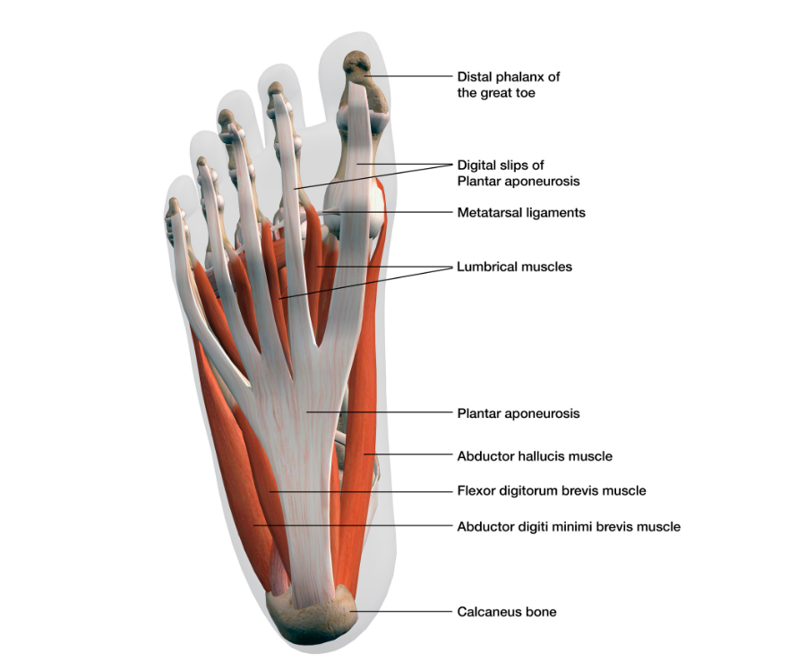
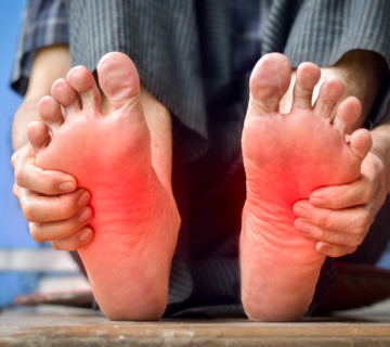
- Ligaments: These tough bands of tissue connect bone to bone. The most famous is the plantar fascia, a thick band that runs along the bottom of the foot from the heel to the toes. When this gets inflamed, it causes a very common and painful condition called plantar fasciitis.
- Tendons: These attach muscles to bones. The Achilles tendon is the largest and strongest tendon in the body, connecting your calf muscles to your heel bone.
- Muscles: Your foot has intrinsic muscles (located entirely within the foot, controlling fine movements of the toes) and extrinsic muscles (originating in the lower leg, providing the power for major movements like pointing your toes or lifting your foot).
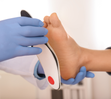
By understanding the anatomy of the human foot, you can better appreciate the complex mechanics required for even a simple stroll around the block—and why finding a shoe with proper arch support is so crucial!

No comment